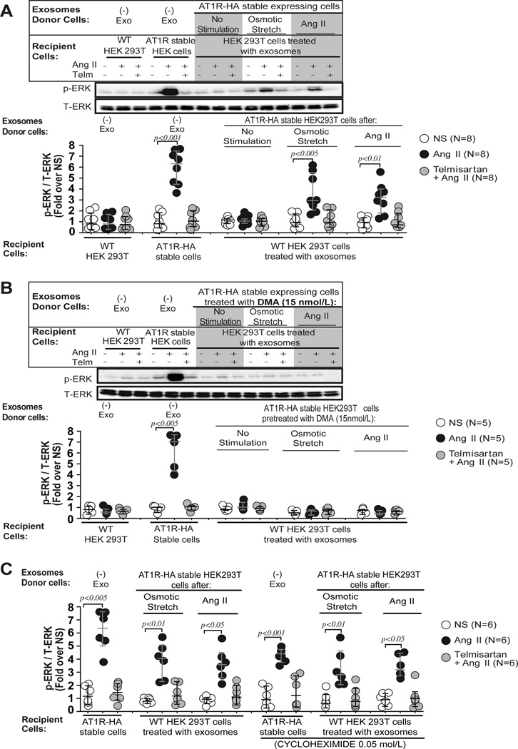
Figure 3.
Exosome derived AT1Rs are able to signal via classical GPCR pathways. A) HEK 293T cells preincubated with exosomes derived from conditioned media of AT1R-HA stable cells after osmotic stretch or AngII stimulation show a significant increase of p-ERK levels after AngII stimulation but not when cells were pre-incubated with exosomes derived from non-stimulated AT1R-HA stable cells. The AngII induced p-ERK response was blocked by the AT1R receptor blocker, Telmisartan (Telm). B) In a separate experiment AT1R-HA stable cells were pretreated with the inhibitor of exosome release, dimethyl amiloride (DMA,15 nmol/L), and exosomes were isolated from conditioned media of cells after stimulation with osmotic stretch or AngII. HEK 293T cells harvested with exosomes derived from AT1R-HA stable cells treated with DMA, failed to respond to AngII stimulation. A–B) Statistical significance was determined by 5 independent Kruskal-Wallis tests with post-hoc Dunn’s test comparing the p-ERK/T-ERK levels of AngII and Telm + AngII to the p-ERK/T-ERK levels of control non-stimulated (NS) in each group of recipient cells. C) HEK 293T cells preincubated with cycloheximide (0.05 mol/L) and exosomes derived from conditioned media of AT1R stable cells after osmotic stretch or AngII stimulation show a significant increase of p-ERK levels after AngII stimulation. Inhibiting protein synthesis with cycloheximide did not result in a reduction in AngII-induced p-ERK levels in recipient cells treated with exosomes. Statistical significance was determined by 6 independent Kruskal-Wallis tests with post-hoc Dunn’s test comparing the p-ERK/T-ERK levels of AngII and Telm + AngII to the p-ERK/T-ERK levels of control non-stimulated (NS) in each group of recipient cells. Additional comparison of the p-ERK/T-ERK levels of preselected pairs of Ang II columns with or without cycloheximide was performed in each 3 type of recipient cells (AT1R-HA stable cells no exo; HEK 293T cells + exo OSM; HEK 293T cells + exo AngII). Data are represented as median with 1st and 3rd quartile.

