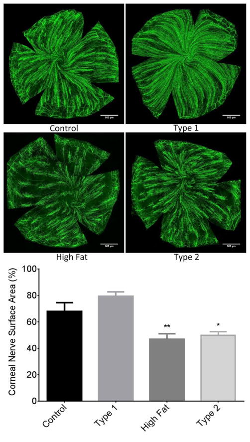
Figure 4. Effect of DIO, type 1 or type 2 diabetes on innervation of sub-epithelial layer of the whole cornea stained with anti-tubulin.
The groups examined and the number of mice in each group was the same as described in Table 1. The images show examples of sub-epithelial layer of corneal nerves of a control mouse (top left), type 1 diabetic mouse (top right), mouse fed a high fat diet (bottom left) and type 2 diabetic mouse (bottom right). Data are presented as the mean ± S.E.M. * p < 0.05 compared to control mice; ** p < 0.01 compared to control mice; one-way analysis of variance with Dunnett’s pairwise test for multiple comparisons. Scale bar = 500 μm.

