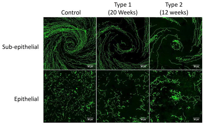
Figure 6. Representative images of sub-epithelial corneal nerves and corneal nerves penetrating the epithelium from control, type 1 diabetic and type 2 diabetic mice.
Corneal nerves of the sub-epithelial layer and penetrating the cornea epithelium from control mice, type 1 diabetic mice (20 weeks duration of hyperglycemia) and type 2 diabetic mice (12 weeks duration of hyperglycemia) were visualized by immunohistochemical staining. Images were collected from the region of the whorl for both the sub-epithelial nerves and nerves penetrating the cornea. Scale bar = 50 μm.

