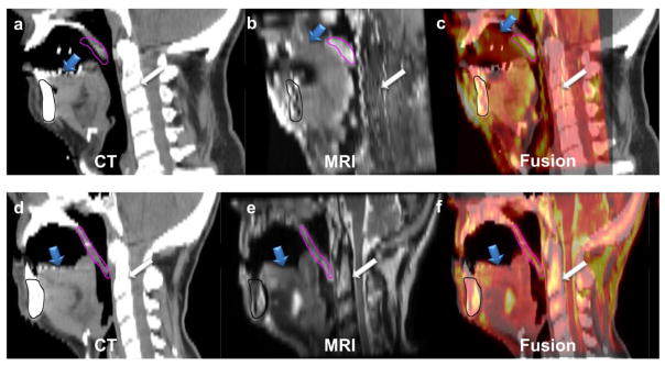
Fig. 4.

The upper panel shows the mid-sagittal sections of radiotherapy-simulation CT (a) registered to standard diagnostic MRI (b) with the fusion overlay (c) of a patient in group 1, while the lower panel shows the mid-sagittal sections of radiotherapy-simulation CT (d) registered to MRI acquired using the studied positioning and immobilization setup (e) with the fusion overlay (f) of a patient in group 2. The registration was done rigidly with priority to oral structures in both cases. It clearly illustrates the error in tongue (blue arrow), soft palate (pink contour segmented on MR and propagated to CT), and spine (white arrow) overlay in the case of group 1 compared to the case of group 2 despite perfect mandibular (black contour segmented on MR and propagated to CT) alignment.
