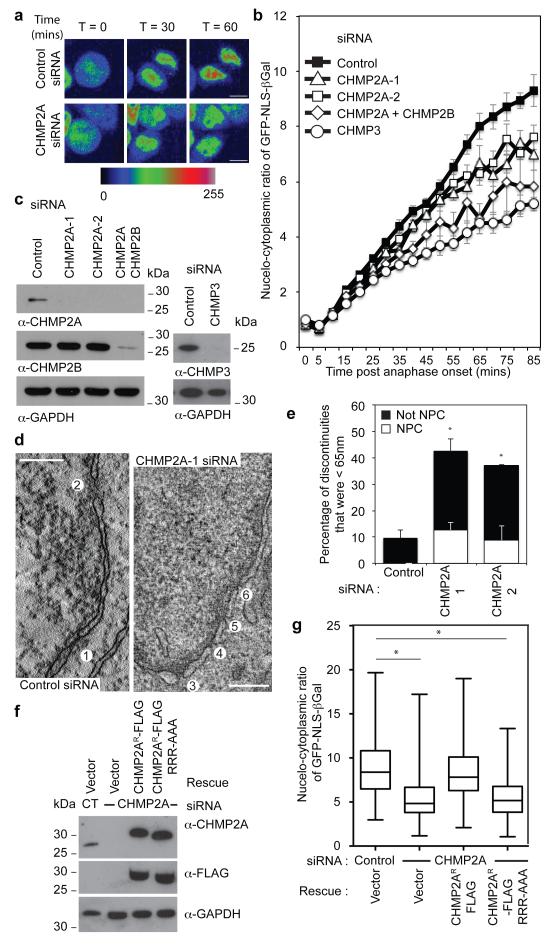
Figure 4. ESCRT-III depletion disrupts nuclear envelope integrity.
A. Timelapse analysis of NE-sealing in siRNA transfected HeLa cells stably expressing H2B-mCh and GFP-NLS-βGal. GFP-signal presented according to pseudocolour scale at the indicated timepoints. Scale bar is 10 μm, a single image was pseudocoloured for demonstrative purposes. B. Quantification of NE-sealing from siRNA treated cells as in A, (Cells were quantified at each time point; Ctrl 140 cells from 7 independent experiments; CHMP2A-1, 98 cells from 5 independent experiments, P = 0.047; CHMP2A-2, 80 cells from 4 independent experiments, P = 0.023; CHMP2A + CHMP2B, 60 cells from 3 independent experiments, P = 0.006; CHMP3, 34 cells from 3 independent experiments, P = 0.002. All values quoted ± S.E.M.; 2-tailed Student’s t-test used to assess significance after 85 minutes). C. Western blotting of cell lysates from B with anti-CHMP2A, anti-CHMP2B anti-CHMP3 or anti-GAPDH antisera. D. Z-slices extracted from a correlative tomographic reconstruction of the NE at 60 minutes post anaphase onset from the indicated siRNA-transfected mCh-Tub HeLa cells. The numbered circles correspond to discontinuities labeled in the 3D reconstructions in Extended Data Figure 10A, scale bar is 200 nm, image representative of 6 (control) and 12 (CHMP2A-1 siRNA) tomographic reconstructions. E. The percentage of discontinuities smaller than 65 nm was scored. Discontinuities in this range that were not NPCs as a percentage of total discontinuities (including NPCs) for n number of reconstructed tomograms: Control 9.4 ± 3.0, n = 6; CHMP2A-1, 29.9 ± 4.7, P = 0.01, n = 12; CHMP2A-2, 28.3 ± 2.0, P = 0.021, n = 2. The increase in the percentage of non-NPC discontinuities was assessed by 2-tailed Student’s T-test (average diameter of non-NPC discontinuities was 38 ± 22 nm (CHMP2A-1) and 58 ± 19 nm (CHMP2A-2)). F. Western blotting of lysates from siRNA-treated HeLa cells stably expressing H2B-mCh, GFP-NLS-βGal and siRNA-resistant CHMP2AR-FLAG with anti-CHMP2A, anti-FLAG or anti-GAPDH antisera. G. Quantification of NE-sealing from cells treated with siRNA as in F and imaged from 4 independent experiments, (Mean nucleo-cytoplasmic ratio given 85 minutes post anaphase onset ± S.D, 2-tailed Student’s T-test was used to assess significance across 4 independent experiments (*); Ctrl, 8.9 ± 3.1, n = 174 ; CHMP2A siRNA 5.4 ± 2.6, n = 171, P = 0.0006; CHMP2A siRNA + CHMP2AR-FLAG 8.4 ± 3.3, n = 132, not-significant; CHMP2A siRNA + CHMP2AR-FLAG RRR-AAA 5.4 ± 2.2, n = 196, P = 0.0001).

