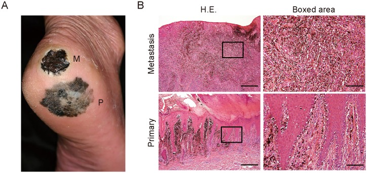
Fig 1. Clinical manifestation and histopathology of the acral lentiginous melanoma patient with wound site metastasis.

(A) Clinical presentation of the right foot of the patient. P, primary lesion; M, metastatic lesion. (B) H&E staining of the primary and metastatic lesions of the patient. The boxed regions are shown at higher magnification in the right panels. Bars: 500 μm (left) or 100 μm (right).
