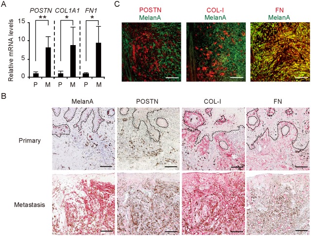
Fig 2. Overexpression of POSTN, COL-1, and FN in the stroma of wound metastasis in a melanoma patient.
(A) Quantitative RT-PCR analysis of POSTN, COL1A1, and FN1 mRNA expression in the primary (n = 3) and metastatic (n = 2) lesions. Data are means ± SD for triplicate samples from one of three representative experiments. *P < 0.05, **P < 0.005. (B) Immunohistochemical staining of the primary and metastatic lesions with antibodies to Melan-A, POSTN, COL-I, and FN. Dashed lines indicate the basement membrane zone. (C) Immunofluorescence analysis of the lower dermis of the metastatic lesion with antibodies to Melan-A (green) as well as those to POSTN, COL-I, or FN (red). FN showed colocalization (yellow) with Melan-A (melanoma cell marker), whereas POSTN and COL-I were localized only in the stroma. (B, C) Bars = 100 μm.

