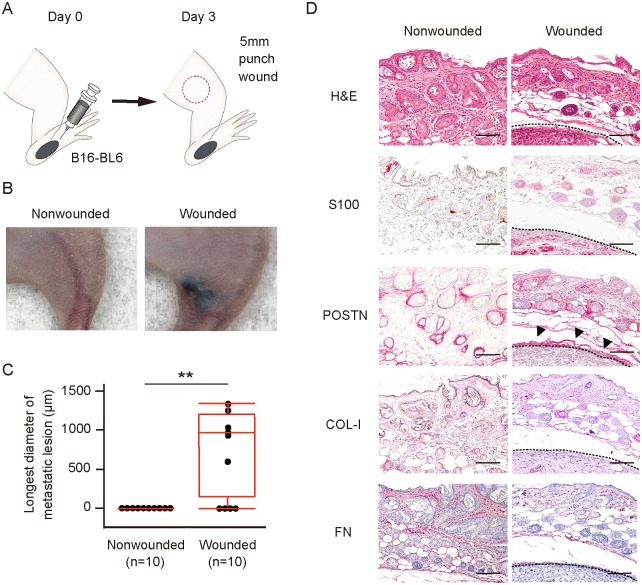
Fig 3. Wound healing process predisposes to metastasis of melanoma in vivo.
(A) Schematic representation of the wound metastasis model. B16-BL6 cells were transplanted into the left hind footpad of a Balb/c nude mouse on day 0, and a 5-mm full-thickness skin wound was inflicted on the left thigh (red circle) on day 3. (B, C) Representative images of (B) and longest diameter of subcutaneous metastasis at (C) the left thigh for wounded and nonwounded (control) mice (n = 10 each) on day 23. Quantitative data are presented as a box-and-whisker plot. **P < 0.005. (D) Representative immunohistochemical staining of S100 (melanoma cell marker), POSTN, COL-I, and FN as well as H&E staining for the left thigh of wounded and nonwounded mice at day 23. Arrowheads indicate POSTN expression surrounding a melanoma tumor cell nest. Dashed lines indicate the periphery of tumor cell nests. Bars = 100 μm.

