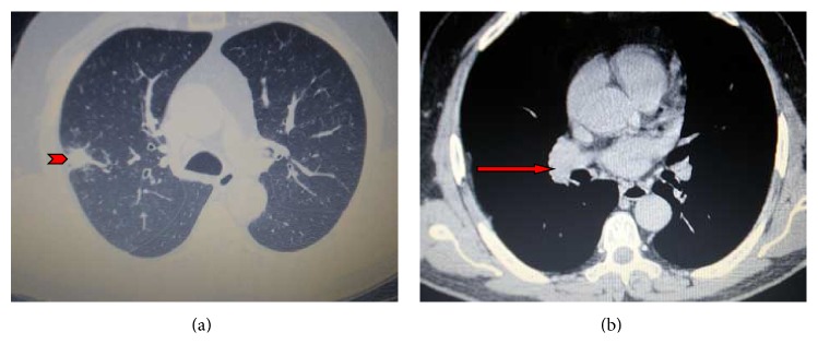
Figure 1.

(a and b) Contrast-enhanced chest computed tomography showing a nodular opacity in the posterior segment of the right upper lobe (arrowhead) accompanied by mild ipsilateral pleural thickening and bilateral mediastinal lymphadenopathy (red arrow).
