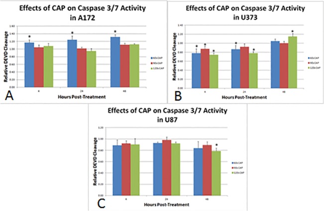
Fig 6. Apoptotic effects of CAP treatment in three different glioma cell lines.

Briefly, cells were treated with CAP for 60, 90 or 120 seconds, incubated with DEVD at 4, 24 and 48 hours post-treatment for 30 minutes, and emission quantified, as per protocol. DEVD cleavage calculated and plotted relative to untreated control cells. (A) Treatment with the lowest dosage of CAP, 60s, induced apoptosis in A172. Higher dosages induced cytotoxicity instead of apoptosis. (B) Apoptosis was significantly increased at 48 hours post-CAP at 120 seconds. At 4 and 24 hours post-CAP, a significant decline in caspase 3/7 activity was detected, likely representing a decline in total population due to the cytotoxic effects observed in Fig 3. (C) No significant increase in caspase 3/7 activity was noted with CAP treatment of U87 cells.* denotes statistical significance compared to control (P <0.05).
