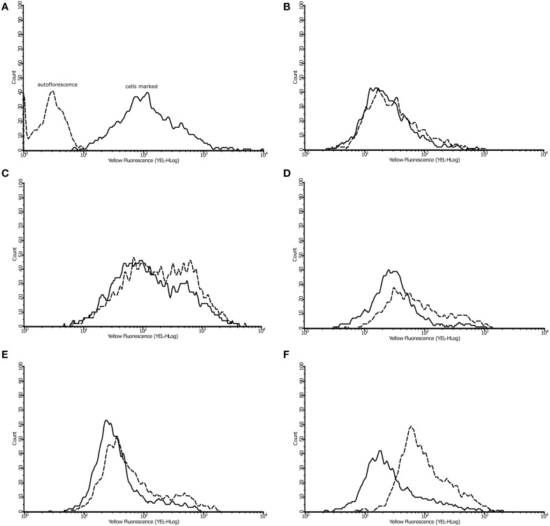
Figure 6.
Effect of argentilactone on intracellular lipid content of P. lutzzi. The presence of lipids was determined by flow cytometry. Cells was stained with dye Nile Red (A). The analysis of yeast cells in presence and absence of argentilactone for (B) 0 h, (C) 6 h, (D) 10 h, (E) 12 h, and (F) 24 h was performed. Line histograms represent the cells treated with argentilactone and dotted histograms represent control cells without treatment.

