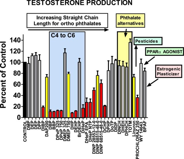
FIG. 1.
Effects of the different in utero maternal treatments on fetal testis testosterone production, collected ex vivo for 3 h incubation (one testis for each of three males per litter, with 3–4 litters per dose group in most cases). Data are expressed as percentage of control from the respective block in which the PE was tested; T Prod data were log10 transformed to correct for heterogeneity of variance. Phthalates are listed from left to right by increasing ester straight side chain length from C2 to C9. Several phthalates which do not have straight side chains from C4 to C6 disrupt fetal testis testosterone production including DIBP, DHeP, DINP, and DCHP. Gray histograms are not significantly different from control (p > 0.10), yellow were equivocal (p ≤ 0.05 to p > 0.01) and red differed significantly (p ≤ 0.01) from the concurrent control value.

