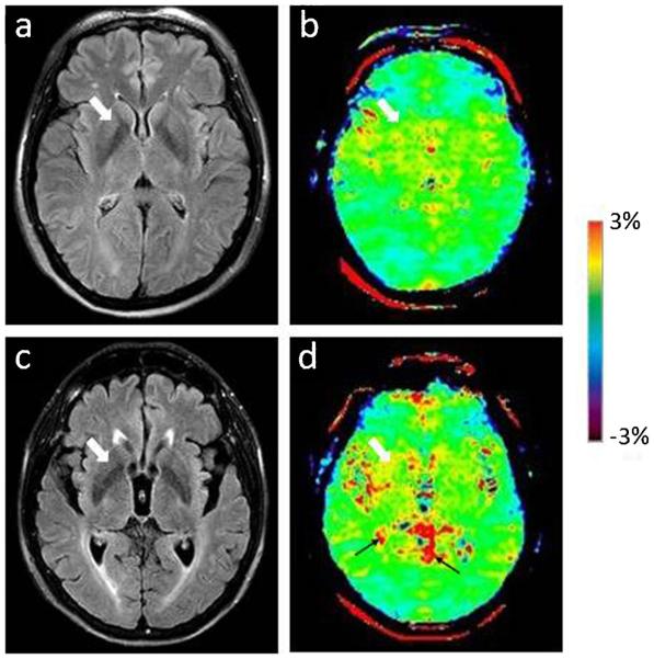
Fig. 3 a.
FLAIR image and b APT-weighted image of a typical normal control (female; 69 years old). c FLAIR image and d APT image of a PD patient (female; 76 years old; H&Y stage 1.5; unified Parkinson’s disease rating scale 48). The CEST/APT imaging acquisition protocol provided B0 inhomogeneity-corrected, APT-weighted images with sufficient signal-to-noise ratios. The APT-weighted intensities in regions of the basal ganglia (white arrow) were higher in PD patients than in normal controls. Note the presence of CSF artefacts (black thin arrows)

