
Figure 3.
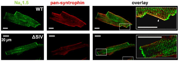
(Upper panels) Isolated mouse ventricular myocyte with double immunofluorescence staining (imaged with confocal microscopy). NaV1.5 (green) is expressed at the IDs, lateral membrane. The punctate staining most-likely represents the expression at the t-tubules. Syntrophin is only expressed at the lateral membrane where it co-localize with NaV1.5 (see arrow in merge showing the yellow region of co-localization). (Lower panels) Stainings of myocytes from genetically-modified mice (truncation of the last three residues of NaV1.5 interacting with syntrophins and SAP97, ΔSIV) illustrating the reduction of Nav1.5 expression exclusively at the lateral membrane, (modified with permission from Shy et al. 2014).