Figure 3.
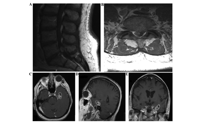
Case 3: Contrast-enhanced magnetic resonance imaging (MRI) of the (A) lumbar spine sagittal and (B) axial sequences reveal intradural spinal metastasis of glioblastoma to the L5-S1 region. Contrast-enhanced MRI of the (C) brain axial, (D) sagittal and (E) coronal sequences show the primary intracranial glioblastoma that was discovered subsequent to the metastatic intradural lumbar spinal lesion.

