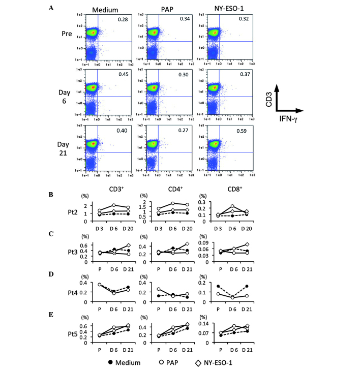
Figure 5.
Detection of tumor antigen-specific T cells. Circulating tumor antigen-specific T cell subsets in patients were characterized by flow-cytometric analysis using immunofluorescent antibody staining for cluster of differentiation 3 (CD3), CD4, CD8 and interferon-γ (IFN-γ). (A) Representative data for CD3 and IFN-γ production in peripheral blood lymphocytes (PBL). PBL were stimulated with prostatic acid phosphatase (PAP) and NY-ESO-1 overlapping peptides. CD3+ IFN-γ+, CD4+ IFN-γ+ and CD8+ IFN-γ+ T cells of (B) patients 2, (C) 3, (D) 4 and (E) 5 are shown, respectively. Each symbol indicated medium (●), PAP (○) and NY-ESO-1 (◇).

