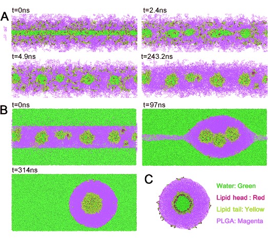
Figure 2.

Snapshots of DPD simulation. A) Formation of water droplets, and assembly of lipids onto the surface of water droplets in the first stage of microfluidic chip. Water is shown in green, lipid head in red, lipid tail in yellow, and PLGA in magenta. B) Formation of RNV in the second stage. C) A slice of the formed water core/PLGA shell/lipid layer RNV.
