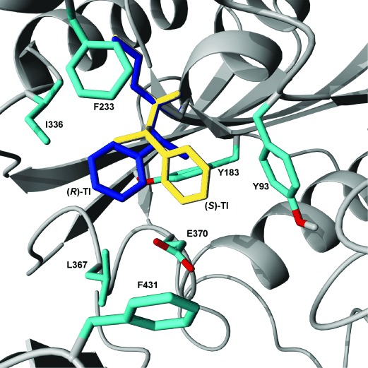
Figure 1.

Close-up view of the active site of CalA. Amino acid residues included in the CalA library (Table 1) are shown as turquoise stick models, while the overall structure is shown as a gray ribbon diagram. Tetrahedral intermediates (TI) of the product, 1-phenylethyl butyrate, are shown in dark blue for the (R)-enantiomer and in yellow for the (S)-enantiomer. The molecular graphics were created by using the YASARA software.[33]
