Figure 4.

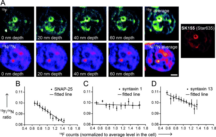
Genetic labeling of proteins and SIMS imaging offer detailed information on how protein metabolism correlates with the presence of a specific protein. A) A series of NanoSIMS images taken at different depth planes in a SK155-labeled cell expressing syntaxin 1. 19F is shown on the upper row; the 15N/14N ratio on the lower row. Inset: for comparison, the fluorescent signal of SK155 in the same cell. Scale bar: 2 μm. B) An analysis of 15N/14N ratios as a function of 19F levels. The number of analyzed cellular regions is 371 for SNAP-25, 281 for syntaxin 1, and 448 for syntaxin 13 samples. A downward trend is observed for all three types of staining, which is statistically significant for SNAP-25 and syntaxin 13 (p<0.01, t-tests), but not for syntaxin 1.
