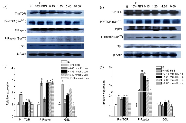
Fig. 2.
Effects of Leu and His on the expression of mTORC1 in CMEC-H
(a) Leu was supplemented for 6 h at the indicated concentrations. mTORC1 levels were determined by immunoblot analysis. Numbers above the lanes refer to the levels (mmol/L) of the supplemented Leu relative to that in the Earle’s balanced salt solution. (b) Densitometric analysis of signals obtained from the mTORC1 immunoblot (a). (c) His was supplemented for 6 h at the indicated concentrations. mTORC1 levels were determined by immunoblot analysis. Numbers above the lanes refer to the levels (mmol/L) of the supplemented His relative to that in the Earle’s balanced salt solution. (d) Densitometric analysis of signals obtained from the mTORC1 immunoblot (c). β-Actin was assessed as a loading control. A representative blot and quantitation of three independent experiments were shown. In all panels, data represent the mean±SD. E: Earle’s balanced salt solution; E+10% FBS: Earle’s balanced salt solution supplemented with 10% fetal bovine serum. Data in the same concentration marked with different letters represent a significant difference (P<0.05)

