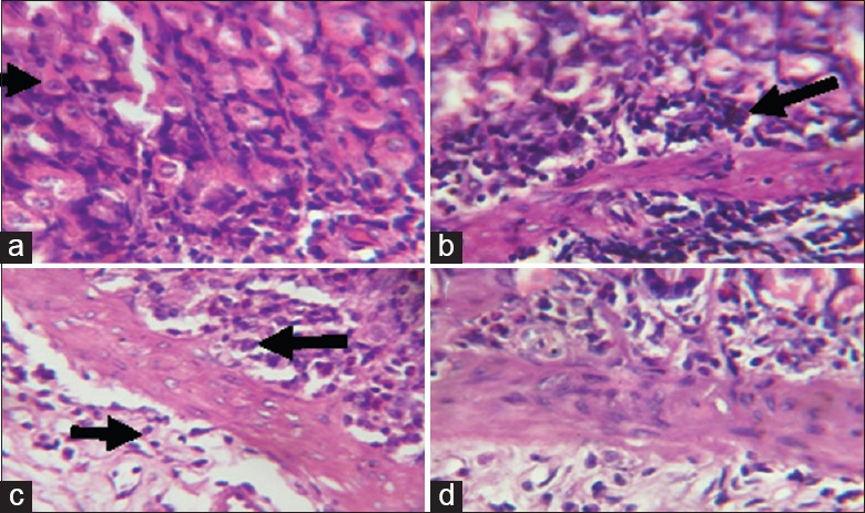
Figure 6.

Photomicrographs showing the gastric mucosa of (a) control rats with predominantly normal parietal cells (black arrow) with only few inflammatory cells; (b) Sodium arsenite-treated rats, showing marked infiltration of the mucosa and submucosa with inflammatory cells; (c) rats pretreated with Kolaviron (KV) 100 mg/kg and (d) rats pretreated with KV 200 mg/kg, both showing only mild infiltration at the base of the mucosa
