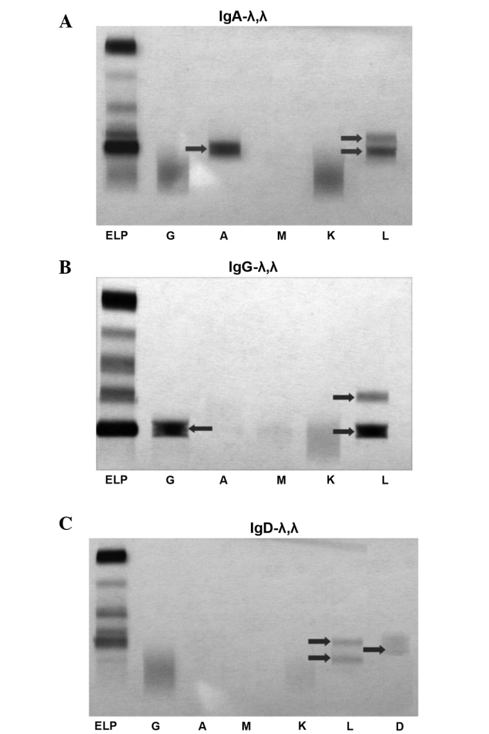
Figure 1.

Two bands for the immunoglobulin (Ig) λ light chain by immunofixation electrophoresis (IFE). (A) IgA-λ,λ, (B) IgG-λ,λ and (C) IgD-λ,λ. Lane ELP, serum protein electrophoresis; lane G, IgG; lane A, IgA; lane M, IgM; lane K, κ light chain; lane L, λ light chain; lane D, IgD.
