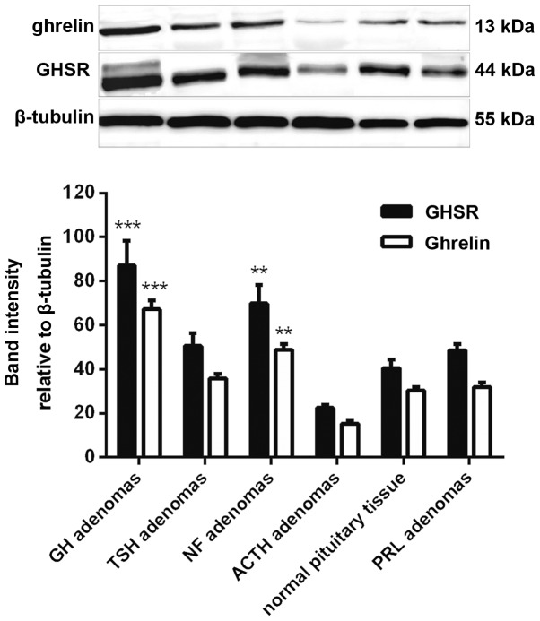
Figure 3.
Western blot analysis showed that the ghrelin and GHSR protein expression followed the same pattern, with the highest mean level in GH adenomas, a moderate level in NF adenomas and the lowest level in ACTH adenomas. **P<0.01 and ***P<0.001 vs. normal pituitary tissue. GH, growth hormone; GHSR, GH secretagogue receptor; PRL, prolactin; NF, non-functioning; ACTH, adrenocorticotropin; TSH, thyroid-stimulating hormone.

