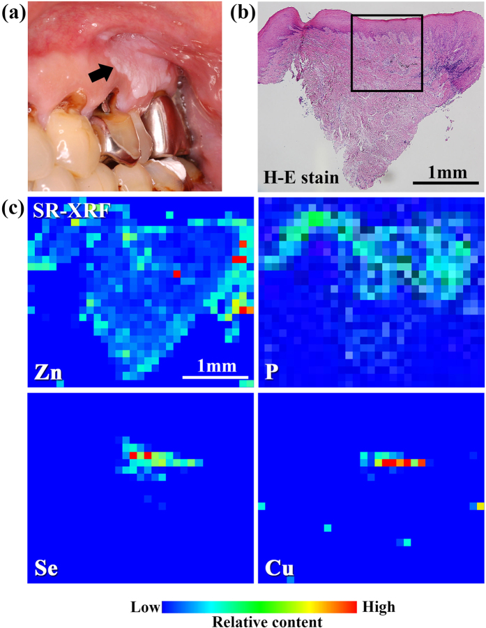
Figure 1. (a) Clinical examination, (b) histopathological image, and (c) SR-XRF elemental distribution images of specimen #7.
(a) Hyperkeratosis was found under the tooth crown (indicated by an arrow). (b) Overlying keratinization, a band-like layer of inflammatory cells, and liquefaction degeneration of the basal cell layer were found. These are the characteristic features of OLP and OLCL. (c) Localisation of Zn, Cu, and Se in the subepithelial layer was observed. P and inflammatory cells were co-localised.

