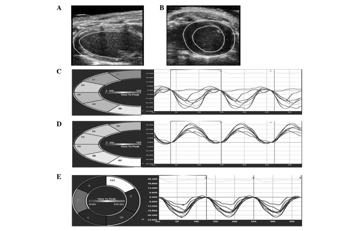
Figure 1.
Representative images of the STE analysis. Endocardium and epicardium were semi-automated and traced by the analyzing software in parasternal (A) long axis view and (B) short axis view. (C) Longitudinal and (D) radial strain value in each segment (including posterior base, mid posterior, posterior apex, anterior base, mid anterior and anterior apex) of left ventricle. (E) Circumferential strain curves for each segment (including anterior free wall, lateral wall, posterior wall, inferior free wall, posterior septum wall and anterior septum wall) of left ventricle. STE, speckle tracking echocardiogram.

