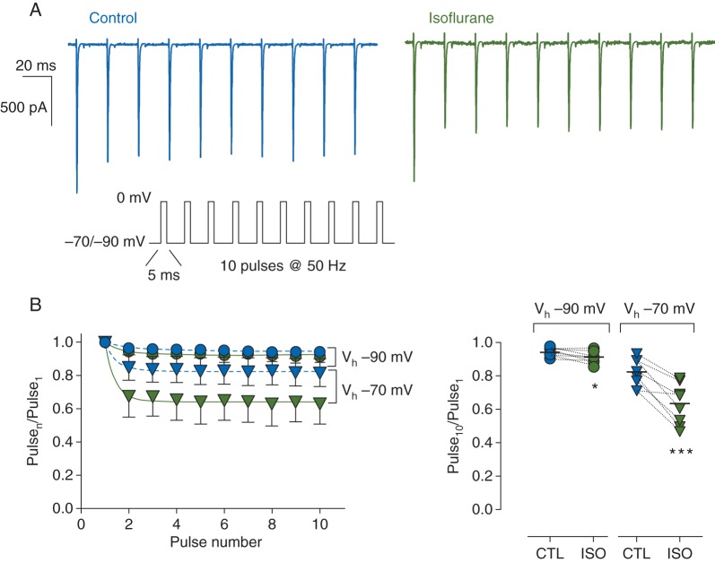
Fig 5.
Effects of isoflurane on peak Na+ current (INa) in isolated rat neurohypophysial nerve terminals during high-frequency stimulation. (a) Representative current traces for a 50 Hz train of 5-ms depolarizing pulses to 0 mV from Vh of −70 mV in the absence (left) or presence (right) of 1.5 MAC isoflurane (protocol in inset). (b) Left panel, shows normalized peak INa [mean (sd), n=8] in the absence (blue symbols) or presence (green symbols) of isoflurane from Vh of −70 mV (triangles) or −90 mV (circles). Peak amplitude of each pulse was normalized to the first pulse (Pulsen/Pulse1) and fitted with a mono-exponential function. Right panel, individual normalized responses for the last pulse (Pulse10/Pulse1) in the absence (blue symbols) or presence (green symbols) of isoflurane. Dotted lines denote paired data. *P< 0.05, ***P<0.001 by two-tailed, paired Student's t-test.

