. 2015 Jun 3;4:e06057. doi: 10.7554/eLife.06057

Figure 1. Purification of the cohesin-loading complex.

(A) Domain organization of Scc2/4. Dotted lines show the Scc2–Scc4 interaction. An arrow indicates the position of a regulated cleavage site (Woodman et al., 2014). (B) Negatively stained Scc2/4 visualized by electron microscopy. Individual particles are shown. (C) Gel filtration chromatograms and SDS-PAGE show that Scc2FL/Scc4 (magenta, left inset) and Scc21–181/Scc4 (purple, right inset) form stable complexes (* marks an Scc2 cleavage product).

DOI: http://dx.doi.org/10.7554/eLife.06057.003

Figure 1.

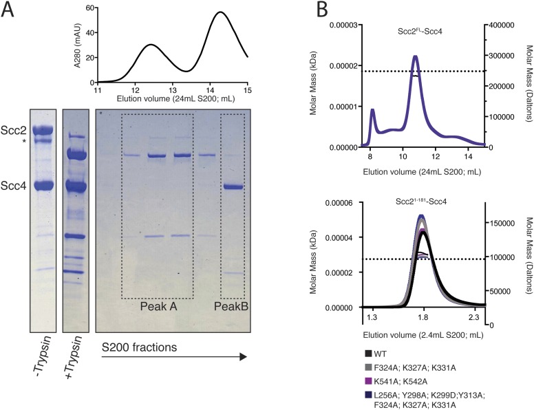
Figure 1—figure supplement 1. Purification and characterization of an Scc2N–Scc4 complex.

Figure 1—figure supplement 1.

(A) Full-length recombinant Scc2/4 was digested with trypsin and analyzed by gel filtration. Fractions from peak B contain full-length Scc4 and the N-terminal 181 or 205 residues of Scc2. A scaled absorbance (A280) trace is shown above for reference. (B) SEC-MALS to determine the molecular weight of Scc2FL-Scc4 (top) and Scc21–181-Scc4 (bottom). Dotted lines show calculated molecular weights for intact complexes. The measured molecular weights were as follows: 238.3 kDa for Scc2FL-Scc4; 104.1 kDa for Scc21–-181-Scc4WT; 100.3 kDa for Scc21–181-Scc4F324A; K327A; K331A; 103.0 kDa for Scc21–181-Scc4K541A; K542A; and 98.8 kDa for Scc21–181-Scc4L256A; Y298A; K299A; Y313A; F324A; K327A; K331A.