
Figure 2.
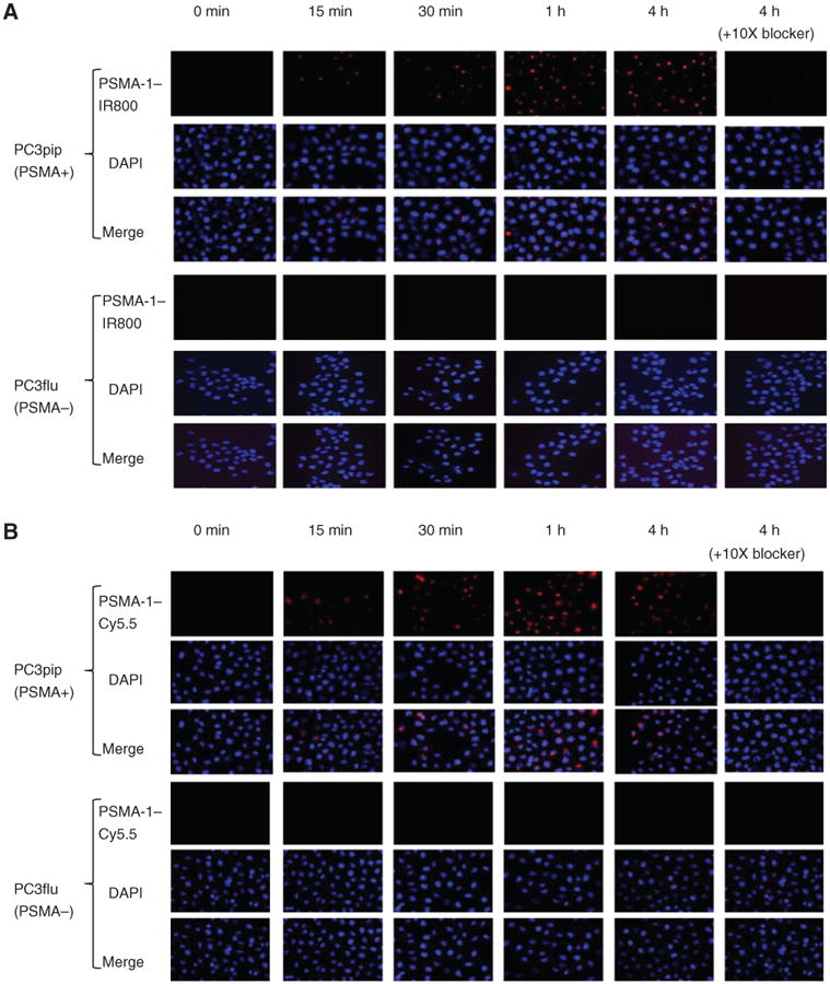
In vitro cellular uptake results of PSMA-1–IR800 and PSMA-1–Cy5.5. PSMA-positive PC3pip cells and PSMA-negative cells PC3flu cells on coverslips were incubated with no probe (0 minutes, A and B) or 1 μmol/L of PSMA-1–IR800 (A) or 1 μmol/L of PSMA-1–Cy5.5 (B) for 15 minutes, 30 minutes, 1 hour, and 4 hours. The nucleus was stained by DAPI (false color blue), and uptake of PSMA-1–IR800 and PSMA-1–Cy5.5 was assessed by fluorescence microscopy (false color red). Specificity of PSMA-1–NIR conjugates to PSMA was evaluated by incubation of PC3pip and PC3flu cells with 1 μmol/L of PSMA-1–NIR conjugates and 10 μmol/L of Cys-CO-Glu, last column in each panel. Signal in PC3pip cells was significantly competed by Cys-CO-Glu, suggesting that the binding of PSMA-1–IR800 and PSMA-1–Cy5.5 to PSMA is specific. Images are taken at 40×. Representative images are shown from 3 independent experiments.