
Figure 5.
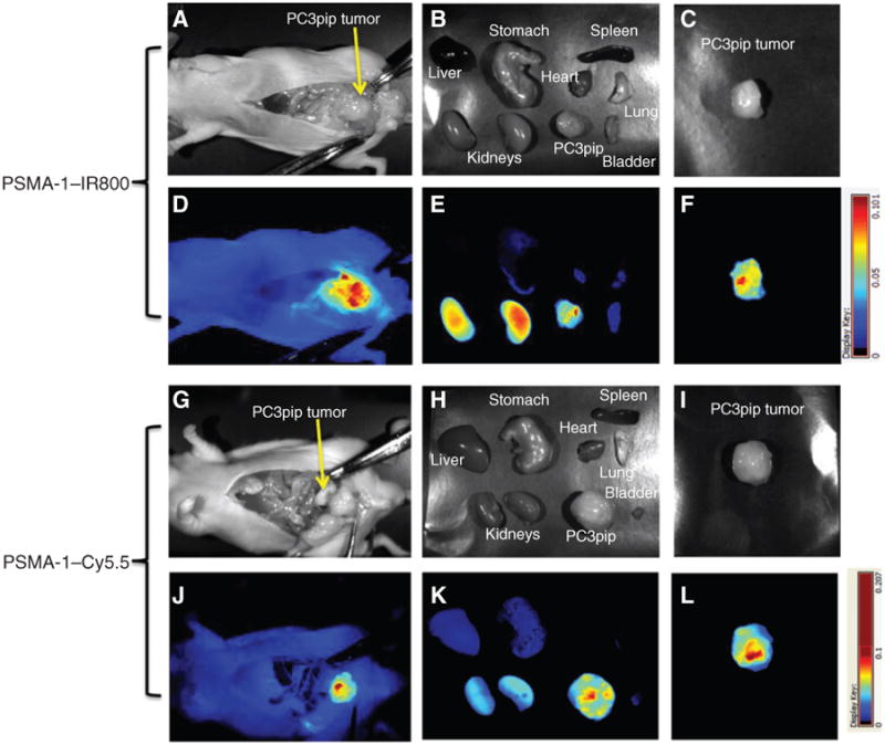
PSMA-1–NIR probes can selectively target orthotopic PSMA-positive PC3pip tumors as shown by Maestro images. Mice received 1 nmol of PSMA–IR800 (A–F) or 1 nmol of PSMA-1–Cy5.5 (G–L) via a tail vein injection. Mice were sacrificed at 4 hours postinjection of PSMA-1–IR800, the abdomen opened to expose the tumor, and both black and white images (A) and fluorescent images (D) were taken. Organs were then harvested for ex vivo images (B and E) and finally tumors were imaged separately ex vivo (C and F). Mice that were administered PSMA-1–Cy5.5 were sacrificed at 24 hours postinjection; the abdomen was opened to expose tumor and then imaged. Both black and white images (G) and fluorescent images (J) were taken, organs were harvested for imaging (H and K), and finally tumors were imaged separately ex vivo (I and L). Pictures are representative images of 4 mice for each probe. Bright fluorescent signal was observed in PC3pip tumor.