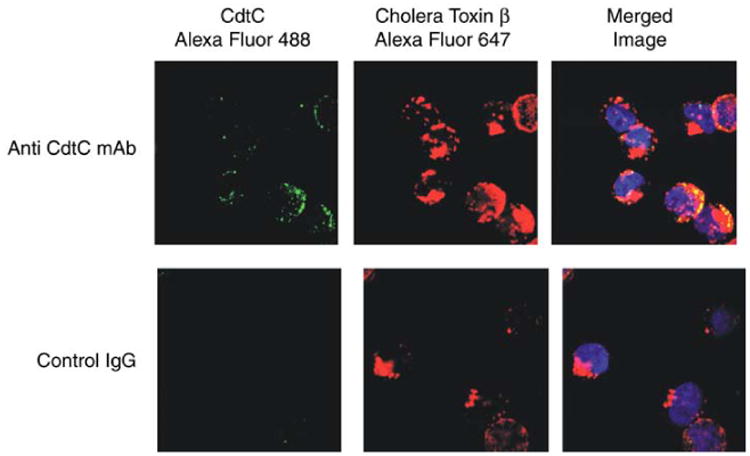
Fig. 3.

Visualization and co-localization of cdt to membrane rafts. To demonstrate that the Cdt subunits localize to membrane lipid rafts, confocal fluorescence microscopy to demonstrate co-localization of the C subunit with the cholera toxin B subunit (CtB) bound to GM1 was used. Jurkat cells were first exposed to CtB-Alexa Fluor 647 for 20 min; the cells were then treated with anti-CtB antisera to induce patch formation. Cells were then exposed to CdtABC for 2 h, washed, and sequentially stained with monoclonal antibody (MAb) to CdtC, goat anti-mouse Ig conjugated to biotin, and streptavidin-Alexa Fluor 488. To control for nonspecific staining, isotype-matched control IgG was used instead of the anti-Cdt MAb. As shown, virtually all fluorescence associated with either CdtC colocalized with GM1 (i.e., CtB fluorescence).
