FIGURE 1:
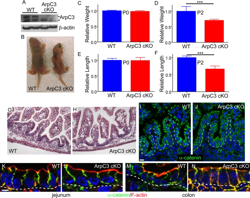
Normal intestinal architecture but failure to thrive upon loss of ArpC3 in the intestinal epithelium. (A) Western blots for ArpC3 and actin in extracts prepared from two WT and two ArpC3 cKO pups. (B) WT and ArpC3 cKO littermates at postnatal day 2. (C–F) Relative weights of P0 and P2 pups, as well as the relative intestinal lengths at these two time points. WT weight and intestinal length was set to 1 in each case. n = 7 or 8, p < 0.0001 for both. (G, H) Hematoxylin and eosin staining of neonatal WT and ArpC3 cKO jejuna. Scale bar, 50 μm. (I, J) α-Catenin (green) and nuclei (blue) in WT and ArpC3 cKO jejuna. Scale bar, 20 μm. Dotted lines indicate basement membrane. (K–N) α-Catenin (green) and F-actin (red) in WT and ArpC3 jejuna (K, L) and colon (M, N). Nuclei are blue; dotted lines indicate the basement membrane. Scale bar, 10 μm.

