FIGURE 2:
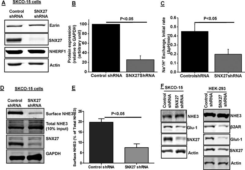
SNX27 regulates NHE3 activity and surface expression in SK-CO15 cells. (A) Western blot analysis of total cell lysate prepared from SK-CO15 cells infected with lentivirus control shRNA or SNX27 shRNA. β-Actin was used as loading control. Ezrin and NHERF1 were used as internal controls. (B) Densitometric analysis of total protein expression from control shRNA and SNX27 shRNA–infected lysates showed that SNX27 protein expression was significantly reduced in SK-CO15 cells containing puromycin as selection marker (n = 4). (C) Na+/H+ exchange was measured in SK-CO15 cells expressing control shRNA or SNX27 shRNA using the pH-sensitive dye BCECF. Results are means ± SE of four separate studies. p values are comparison between control shRNA and SNX27 shRNA. Initial rates are shown. (D) Single study of NHE3 surface expression using biotinylation in SK-CO15 cells. Three dilutions of total cell lysate (10, 20, 40 μl) and avidin precipitate/surface (10, 20, 40 μl) were loaded on SDS–PAGE, and NHE3, SNX27, and GAPDH were identified by immunoblotting; percentage of surface to total was calculated as explained in Materials and Methods. To illustrate the results, in this example, we show results of the loading of one equal amount of total cell lysate (40 μl) and avidin precipitate/surface (40 μl) on SDS–PAGE with NHE3, SNX27, and GAPDH identified by immunoblotting. (E) Quantification presents surface NHE3 normalized to total NHE3 for control shRNA and SNX27 shRNA. Results are means ± SE of four separate studies. p values are comparison between control shRNA and SNX27 shRNA. (F) Western blot analysis of total cell lysate prepared from SK-CO15 and HEK293 cells infected with SNX27 shRNA or control shRNA. Expression of NHE3, Glut-1, and β2AR is shown. β-Actin was used as loading control. A representative result from three independent experiments is shown.

