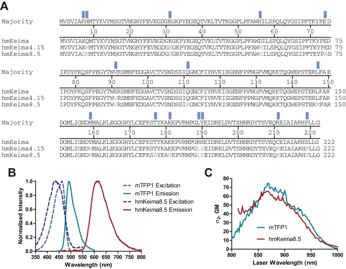
FIGURE 1:
Evolved variants of the long Stokes shift fluorescent protein mKeima. (A) Alignment of mKeima variants. (B) Normalized absorption (dashed lines) and fluorescence emission (solid lines) spectra of hmKeima8.5 (dark blue/red) and mTFP1 (blue/teal). (C) The two-photon cross sections of mTFP1 and hmKeima8.5.

