Figure 1.

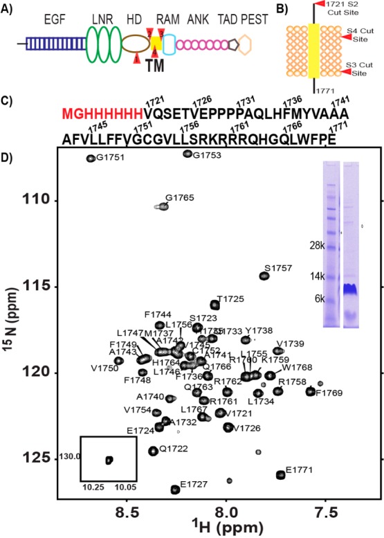
(A) Domain organization of full length Notch1. Sites of proteolysis are colored red. (B) Notch1 TM/JM segment with proteolysis sites colored red. S3 and S4 are γ-secretase cut sites. (C) Sequence of the Notch1 TM/JM segment. (D) Assigned 900 MHz 15N TROSY-HSQC spectrum of the Notch1 TM segment in 15% DMPC/DH6PC bicelles (q = 0.33) at pH 5.5 and 318 K. Backbone amide 1H–15N peaks have been assigned for all of the non-proline residues. The NMR sample included 2 mM DTT, 10% D2O, and 1 mM EDTA. A sodium dodecyl sulfate–polyacrylamide gel of the NMR sample and the single indole side chain 1H–15N peak are shown in the insets.
