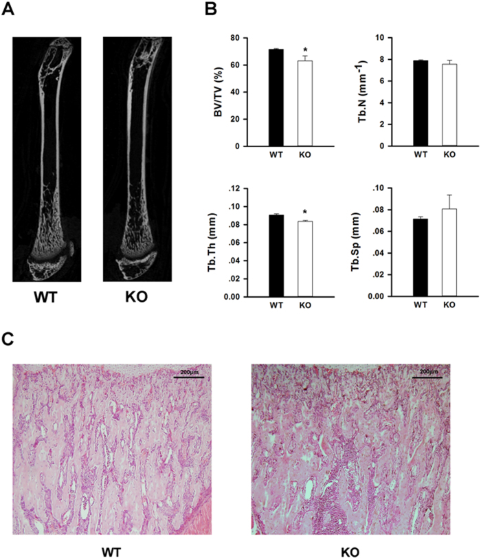
Figure 4.
Deletion of IGF1R in MSCs resulted in decreased bone mass. A: Representative microcomputed tomography (μCT) image from wildtype (wt) and MSC-Igf1r−/− (ko) mice in 7-week-old female mice. B: Quantitative analysis of μCT parameters of trabecular bone in distal femoral metaphysis in wt and ko female mice: bone volume/tissue volume (BV/TV), trabecular number (Tb.N), trabecular thickness (Tb. Th), and trabecular spacing (Tb. Sp). Data presented as mean±SD (n=5, ∗P<0.05). C: Representative H&E staining of tibiae from wt and ko mice. Scale bar=200 μm.

