Figure 1.
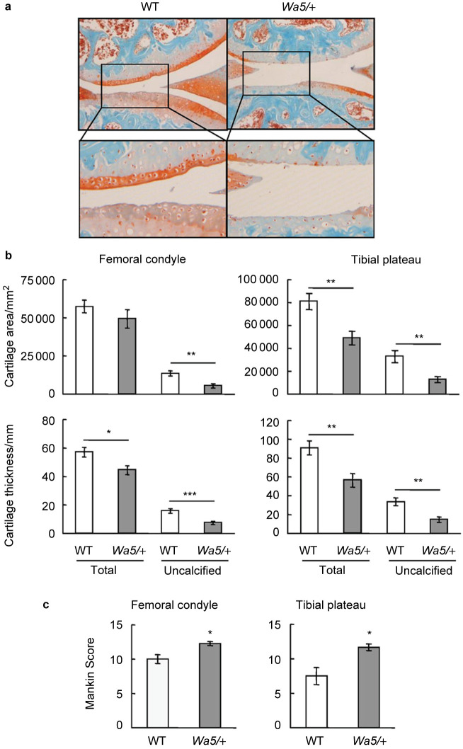
EgfrWa5/+ mice exhibit accelerated osteoarthritis progression after DMM surgery. (a) Representative Safranin O/Fast Green staining images of mouse knee joints show increased articular cartilage degradation in EgfrWa5/+ mice 3 months after DMM surgery. Bottom panels are magnified images of top panels. (b) Total articular cartilage, uncalcified articular cartilage, and their respective thickness were quantified at both femoral condyle and tibial plateau regions. (c) Mankin score graded by blinded observers confirmed more articular cartilage destruction in EgfrWa5/+ mice (n=9) compared to WT (n=10) mice after DMM surgery. * P<0.05; ** P<0.01; *** P<0.001.

