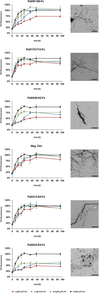
Figure 6.

Influence of scFv-Fc antibodies (Yumabs) on Aβ42 fibrillogenesis. Left: 5 μM Aβ42 monomers were incubated with 2 μM (red), 1 μM (blue), 0.5 μM (green) or 0 μM (black) of scFv-Fc antibodies at 37°C under constant agitation of 300 rpm, ThT fluorescence was monitored over a time course of 96 h. All measurements were carried out in triplicates, the error bars represent the respective standard deviation. Right: representative TEM images of the fibrils formed from of 5 μM Aβ42 monomer after 96 h incubation in the presence of 2 μM scFv-Fc antibody, scale bar corresponds to 400 nm.
