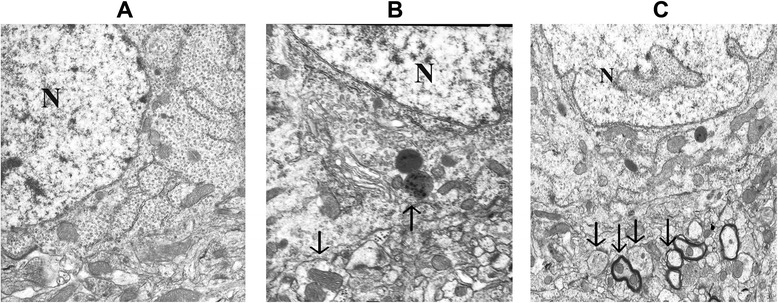
Fig. 2.

Electron micrographs of the hippocampus detected at 12 h following sham operation (a), CLP-12 h (b), and CLP + PDTC-12 h (c). a Sham-operated control rats showed organelles almost without pathological changes; no alteration of tissue integrity could be observed in low magnification images. Magnification: ×10,000. b A large autophagosome contains mitochondria and other organelles; endoplasmic reticulum matrix into adjacent lysosomal structures (arrow). Magnification: ×15,000. c CLP + PDTC-12 h displaying multiple double or multiple-membrane autophagic vesicles (arrows) in the cytoplasm, with loss of discernable organellar fragments; autophagosomes assume a more complex appearance, with redundant whorls of membrane-derived material. Magnification: ×10,000. CLP cecal ligation and puncture, PDTC pyrrolidine dithiocarbamate
