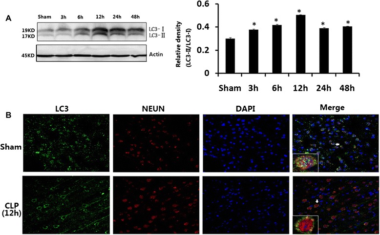
Fig. 3.

a LC3 in brains harvested 3, 6, 12, 24, and 48 h after CLP. b Immunofluorescence for LC3 in neurons after CLP injury. Values of protein data were related to signals obtained from actin protein and given as relative arbitrary units. LC3-II significantly increased in CLP rats at 6, 12, 24, and 48 h after surgery compared with sham-operated rats. Three-color staining for anti-LC3 antibody (green), NeuN (red), and DAPI (blue) showed that a staining pattern changes from largely diffuse to predominantly punctate and cytoplasmic in hippocampal neurons after CLP. Data were expressed as mean ± SEM, n = 6/group. *P < 0.05 vs sham-operated group. LC-3 microtubule-associated protein light chain-3, CLP cecal ligation and puncture, DAPI 4,6-diamidino-2-phenylindole
