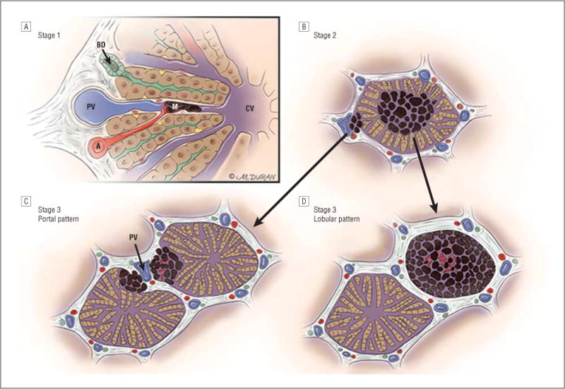
Figure 7.

Progression of ocular melanoma metastasis to the liver through stages 1, 2, and 3. A, In stage 1, up to 50 μm in diameter of avascular aggregates of melanoma (M) arise in sinusoidal spaces via the hepatic arteriole (A); blood flows in the sinusoidal space from the hepatic arteriole and portal venule (PV) toward the central vein (CV); bile flows in the opposite direction toward a bile ductule (BD). B, In stage 2, aggregates of melanoma coalesce to form 51 to 500 μm in diameter of metastases and co-opt the portal venule or infiltrate the hepatic lobule with loss of hepatocytes, proliferation of stellate cells, and the appearance of pseudosinusoidal spaces. C, In stage 3, metastases that measure greater than 500 μm in diameter may grow in a portal pattern in which they co-opt the portal venule (PV). D, Metastases may also infiltrate the hepatic lobule in the lobular pattern.
