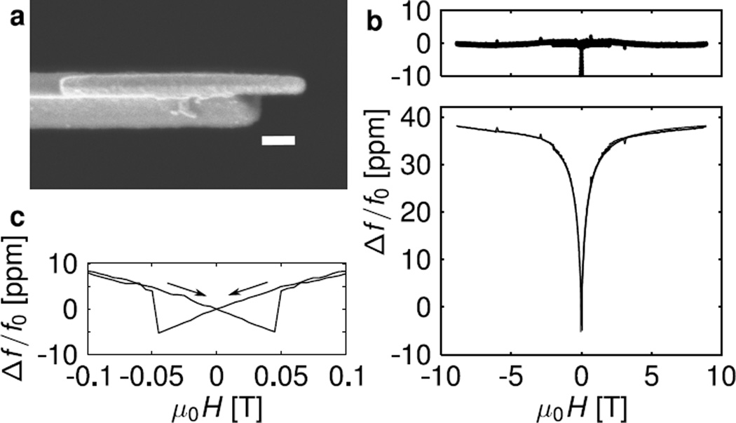
Figure 4.
(a) Scanning electron micrograph of the cantilever’s leading edge, showing a nickel nanorod tip overhanging the cantilever’s leading edge by 350 nm (scale bar = 200 nm). (b) Characterization of the cantilever tip’s nickel nanorod by frequency-shift cantilever magnetometry. Fractional cantilever frequency shift versus field in parts per million (middle; solid line), the best-fit curve (middle; dotted line, indistinguishable from the data), and fit residuals (top). (c) Expanded view of the observed frequency shift near zero field, showing hysteresis.

