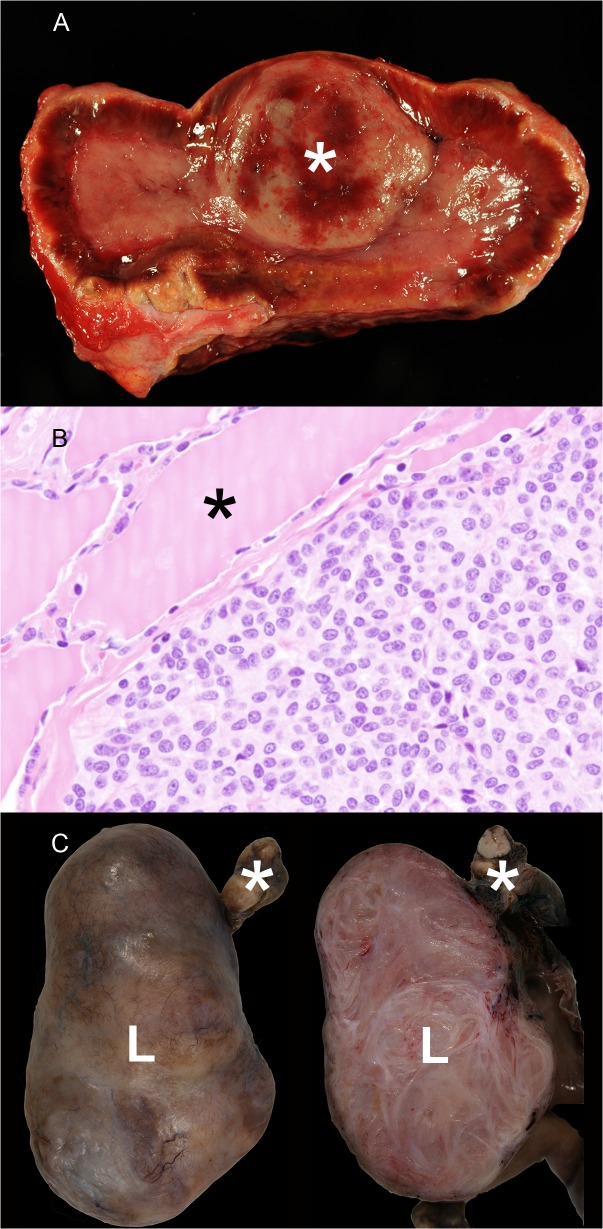
Fig 3. Endocrine and genital neoplasia in wild felids.
A) Adrenal gland, tiger, 19 years, male (animal no 27). Unilateral pheochromocytoma (asterisk). B) Parathyroid gland, leopard, 9 years, female (animal no. 7). Parathyroid gland adenoma characterized by a solid growth pattern. The parathyroid gland adenoma displayed a capsule and compresses adjacent normal follicles of the thyroid gland (asterisk). H&E-staining. C) Ovary, leopard, 17 years, female (animal no. 13). The left part of the picture shows an encapsulated, well demarcated leiomyoma (L) attached to normal ovary tissue (asterisk). The right part of the picture demonstrates the firm, nodular cut surface of the leiomyoma (L) that is characterized by irregularly arranged interwoven tissue bundles.

