Figure 1.

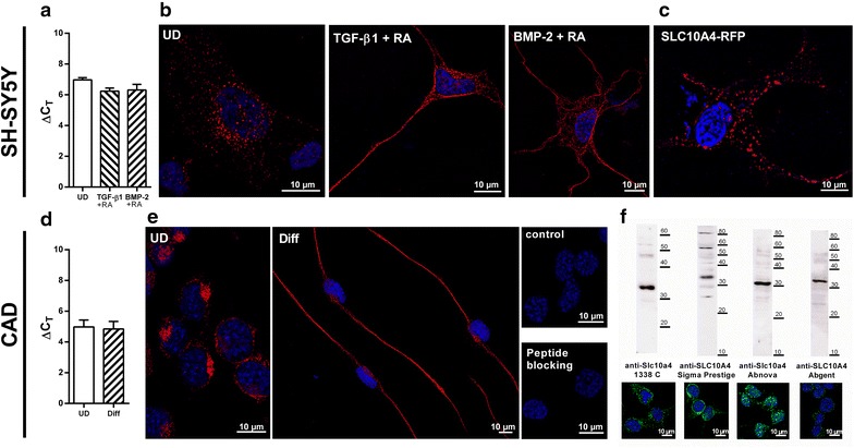
Expression and subcellular localization of SLC10A4 in SH-SY5Y and CAD cells. a Relative SLC10A4 gene expression in SH-SY5Y cells after differentiation with TGF-ß1 + RA or BMP-2 + RA. Values represent mean ± SD of triplicate measurements. b Immunofluorescence analysis of the subcellular expression of the SLC10A4 protein in SH-SY5Y cells. Cells were either untreated (UD), or were differentiated with TGF-ß1 + RA or BMP-2 + RA over 4 days prior to immunolabeling. The SLC10A4 protein was detected with the anti-Slc10a4 1338 C antibody (1:1,000) and the Cy3-labelled anti-rabbit secondary antibody (1:800, red fluorescence) and nuclei were stained with DAPI (blue fluorescence). In all cases, the SLC10A4 protein showed a vesicle-like expression pattern within the perikarya and along the neurite-like cellular protrusions. c Even when a fluorescence-tagged SLC10A4-RFP construct was transiently transfected into SH-SY5Y cells, the SLC10A4-RFP protein showed a clear vesicular sorting pattern. d Relative Slc10a4 gene expression analysis in differentiated (Diff) and undifferentiated (UD) CAD cells. The values represent mean ± SD of triplicate measurements. e Endogenous expression of the SLC10A4 protein in CAD cells, cultivated in FCS containing medium (UD) or FCS-free medium (Diff). The SLC10A4 protein was detected with the anti-Slc10a4 1338 C antibody (1:500) and the Cy3-labelled secondary antibody (1:800, red fluorescence). For control, the primary anti-Slc10a4 antibody was omitted (control) or the antibody was pre-incubated with the immunizing peptide (peptide blocking). f Immunofluorescence detection of the SLC10A4 protein was performed with different SLC10A4-directed antibodies (green fluorescence): self-generated polyclonal rabbit anti-Slc10a4 1338 C antibody, rabbit anti-SLC10A4 Sigma Prestige antibody, rabbit anti-Slc10a4 Abnova antibody, and rabbit anti-SLC10A4 Abgent antibody. Membrane protein enriched fractions of the CAD cells were also subjected to Western Blot analysis with the same antibodies and revealed specific bands for the SLC10A4 protein at an apparent molecular weight of 30–32 kDa.
