
Fig. 1.
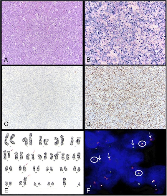
Representative case of plasmablastic lymphoma. a Neoplastic cells have plasmablastic morphology, with a prominent nucleolus and moderate amount of cytoplasm. Mitotic figures and tingible-body macrophages are abundant and impart a “starry-sky” pattern (H&E, ×200). b The neoplastic cells are diffusely positive for EBV-encoded RNA (EBER) by colorimetric in situ hybridization (×200). c CD20 expression is absent. This case was negative for CD19 and positive for CD38 by flow cytometry (data not shown) (×200). d MYC overexpression is positive by immunohistochemistry (×200). e Karyotype of case 30 (nasopharyngeal mass): 46, XY, del(6)(q23q29),t(8;14)(q24;q32), add(20)(p13). f Fluorescence in situ hybridization using a dual-color break-apart probe specific for the MYC locus on formalin-fixed paraffin-embedded tissue (case 30) showing split signals in ~80 % of nuclei (circle: fusion signal; arrow: split signal)