
Figure 2.
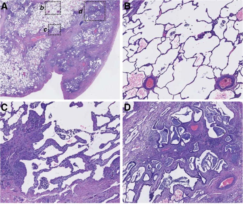
Spatial heterogeneity of pathophysiology in a single representative IPF biopsy.
A) Low-power magnification of an H&E-stained paraffin section of explanted lung tissue from an IPF patient. Pleural surface is at lower right. b, c, and d indicate regions shown at higher magnification in: (B), normal-appearing alveolar structure, (C) transition zone underlying normal-appearing parenchyma with thickened alveolar walls, (D) advanced scar tissue with microscopic honeycombing and bronchiolization.