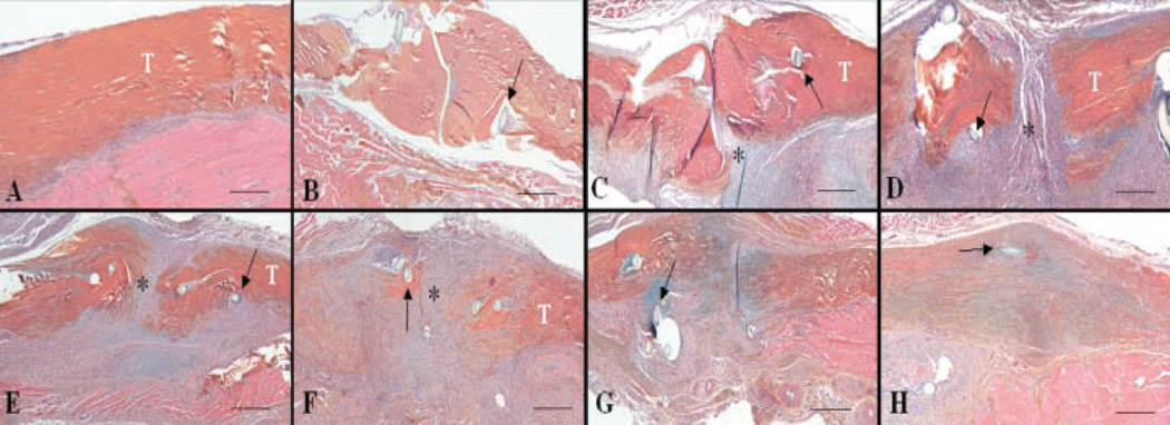
Figure 1.
Representative histology sections of normal (A) and repair FDL tendon at days 3 (B), 7 (C), 10 (D), 14 (E), 21 (F), 28 (G), and 35 (H) post-repair (4×). Sections were stained with Alcian Blue/Hematoxylin and OrangeG. Of note is the fibroblastic granulation tissue (*) that fills in the repair site between tendon ends (marked as T) (C–F) and is progressively remodeled with increasingly organized collagen fibers (F–H). Sutures are marked with arrows. Scale bars represent 25 µm.

