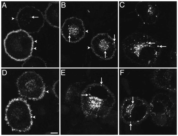
Fig. 1.
Localization of the MOR in KNRK-MOR cells. Cells were unstimulated (A), incubated with 1 μM endomorphin-1 for 10 min (B) or 30 min (C), or 30 min with 1 μM naloxone (D), or incubated with 1 μM endomorphin-2 for 30 min (E) or 1 μM DAMGO for 30 min (F). The MOR was detected using FLAG M1 (A, C, E) or MOR384–398 (B, D, F) antibodies. Images are single optical sections. In unstimulated cells, the MOR was present at the plasma membrane (arrowheads) and in a few endosomes (arrow). After incubation with agonists, the MOR was present in many superficial and perinuclear endosomes (arrows), and there was diminished surface immunoreactivity. Cells illustrated in A are all stained, but with different intensity. This reflects variability in staining seen in transfected cells. Naloxone caused retention of the MOR at the plasma membrane (arrowheads).
Scale bar = 5 μm.

