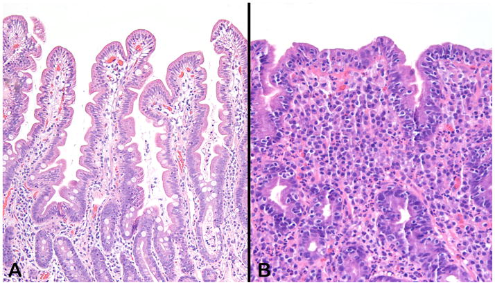
Figure 1.
Sections from normal duodenal mucosa (A) and from a mucosal biopsy with coeliac disease (B). In contrast to the long villi with only minimal numbers of intraepithelial lymphocytes, panel B shows an epithelium studded with lymphocytes and a lamina propria obliterated by a mixed inflammatory infiltrate consisting of lymphocytes, plasma cells, eosinophils, and rare neutrophils. The normal mucin content of the normal goblet cells, evident in panel A, is completely depleted in the mucosa depicted in panel B. Both sections were stained with hematoxylin and eosin and photographed at an original magnification of 10X.

