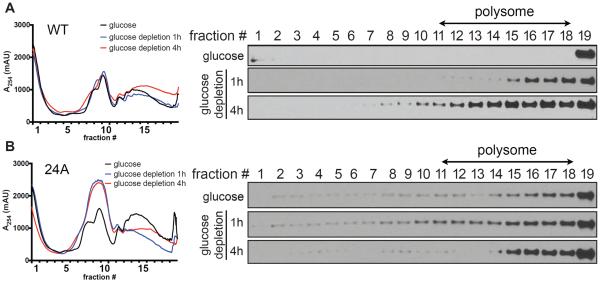
Figure 3. Puf3p associates with polysomes following glucose depletion.
(A) Puf3p WT polysome profiles in glucose or following glucose depletion for 1 h or 4 h.
(B) Puf3p(24A) polysome profiles in glucose or following glucose depletion for 1 h or 4 h. Polysomes were fractionated by sucrose gradients (7–47%, w/v). Samples were subjected to continuous A254 measurements and separated into 19 fractions. Proteins from each fraction were precipitated by TCA and analyzed by Western blot. Note that Puf3p associates with polysomes only following glucose depletion, whereas Puf3p(24A) is mislocalized to all fractions regardless of glucose availability.

