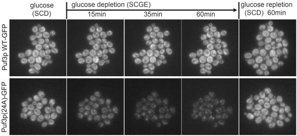
Figure 5. Puf3p(24A) phosphomutant forms punctate foci following glucose depletion.
Live cell imaging of cells expressing Puf3p-GFP or Puf3p(24A)-GFP using the CellASIC microfluidics platform, before and after switch to glucose depletion medium for the indicated times. Puf3p-GFP was uniformly distributed in the cytosol and not specifically localized to mitochondria regardless of glucose availability. Strikingly, Puf3p(24A) forms foci only after switch to glucose depletion medium. Approximately 40% of cells expressing Puf3p(24A)-GFP exhibited foci following glucose depletion, in contrast to 0% of cells expressing WT Puf3p-GFP. See also Figure S5 and Supplementary Videos. These foci exhibited partial co-localization with the p-bodies marker Dcp2p (Figure S5). Both Puf3p(24A) and Dcp2p formed foci only after switch to glucose depletion medium (Figure S5).

